An Irish scientist hailing from Donegal town will light up Times Square New York next month with his striking image of a lung cancer cell surrounded by a green glow.
The image was taken by Dr Martin Barr, Clinical Scientist and Adjunct Assistant Professor at the Institute of Molecular Medicine in Trinity College Dublin and St James’s Hospital, who was today announced as a winner of an international competition that showcases the beauty of cells and the inspiring research conducted by cellular biologists around the world.
The three winners of GE Healthcare Life Sciences’ 2013 Cell Imaging Competition will see their images displayed on high resolution screens in New York’s Times Square next month.
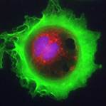
Dr Barr’s image which was taken as part of his ongoing research to better understand and fight resistance to lung cancer treatments shows in extraordinary detail a lung cancer cell from one of the most common forms of lung cancer; non-small cell lung cancer (NSCLC).
The cell measures just one thousandth of a millimetre, similar in width to a cotton fibre, and is shown in a low-oxygen environment known as ‘hypoxia’. This environment, commonly seen in many solid tumours, makes cancer cells more resistant to chemotherapy and encourages the cancer to spread in the majority of patients. The aim of Dr Barr’s research is to target cellular processes triggered by hypoxia in order to make tumours more susceptible to chemotherapy.
Lung cancer is the fourth most common cancer in Ireland and is the commonest cause of cancer death in both sexes, where it accounts for 20% of all cancer deaths. In 2013 there were 2,165 new cases of lung cancer in men and women in Ireland with incidence rates for women in Ireland 55% higher than the EU average.
Speaking about what being a winner of this competition means to him and for cancer awareness, Dr Barr said: “As a winner of the GE Healthcare 2013 Cell Imaging Competition, I am absolutely delighted to have one of my images recognised as a prize-winning image by an expert international panel of judges and a public vote of over 23,000 votes.
Lung cancer mortality remains significantly high worldwide and in Ireland continues to increase, particularly in women. While novel strategies to target the various cellular processes implicated in resistance to current therapies unfold, a visual image of a cell can have more of an impact and in some instances speak more loudly than words, and highlights the cellular complexity of a cancer cell.”
“To see my winning image displayed on the large high-definition screens in Times Square in New York is a unique, once-in-a-life time opportunity and something I would never have imagined in my career as a cancer research scientist. The projection of my image in this major international hot-spot will hopefully bring further awareness of lung cancer to the general public.”
The image which won the High-Content Analysis category of the competition was taken using an IN Cell Analyzer, one of GE’s high resolution cell imaging and analysis systems. This technology allows researchers to acquire and analyse multiple images of thousands of cells in a short period of time to aid in the development of tailored drug therapies to combat diseases such as cancer.
Eric Roman, General Manager of Research and Applied Markets, GE Healthcare Life Sciences, said: “This year’s three winning images are once again incredibly beautiful and compelling, reminding us of the cellular complexity behind disease and why the study of cells is so important. We were delighted to receive so many outstanding entries to the competition, which highlights how cell imaging is helping scientists explore the universe of the cell, and so advance our understanding of so many life-threatening and life-limiting diseases. I’d like to thank all the contestants for sending us their images, the judging panel and everyone who cast a vote.”
For seven years, GE’s annual competition has showcased the beauty of cells and the inspiring research of cellular biologists from around the world. This year’s competition attracted over 100 entries from scientists who are using either high-content analysis or high- and super resolution microscopy to investigate at the cellular level a wide variety of diseases such as cancer, muscle disease and the effects of parasitic infections.
- Thu, 25 Apr 2024
- (+353) 07491 25000
- (+353) 086 60 25000